Today, we will continue to talk about experimental equipment of the Frank Laboratory of Neutron Physics complementing the set of neutron devices. In issue #14 of the Newspaper, we introduced to readers the X-ray diffractometer. Presenting the second facility, the atomic force microscope, Head of the Department of Neutron Investigations of Condensed Matter (DNICM) of FLNP D. P. Kozlenko said: “While an x-ray facility helps to investigate crystal materials, atomic force microscopy is helpful in the characterization of nanosystems in both solid and liquid states. Thanks to this method, it is possible to evaluate characteristic dimensions of nanoparticles and then use this data to interpret the results of small-angle neutron scattering.
Responsible for the facility, DNICM FLNP Researcher Yu. E. Gorshkova (in the photo) introduced to me all the opportunities of the atomic force microscope placed in the recently repaired premises: “Since 2012, when the IBR-2 reactor was brought into the regular operation mode again after modernization and was used for a physical experiment, the FLNP user programme was resumed. The number of users of the IBR-2 increases annually. The equipment set is supplemented according to the needs of our team, Russian and foreign partners from the JINR Member States, in order to solve scientific tasks. Our core principle when choosing expensive equipment is the opportunity to use it for a wide range of tasks in the frames of the theme “Investigations of Condensed Matter by Modern Neutron Scattering Methods”.
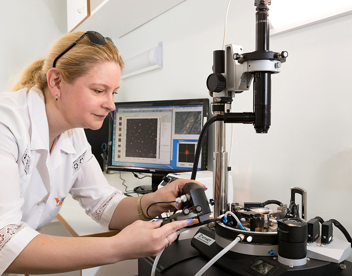
Nowadays, one of the most needed devices is the atomic force microscope produced by the NT-MDT Spectrum Instruments (Zelenograd, Russia). It is made according to the modular design, and one and a half year ago, we bought it in its basic configuration that allows investigation of the topology of the samples’ surface in both contact and non-contact modes sensitive to various interactions of the detector with the surface. It provides an opportunity to measure some characteristics of the surface while scanning, such as viscosity and elasticity spread, electric and magnetic domains.
As for the development of this device, an additional system for the study of samples in a so-called gas-liquid cell was bought in 2019.
Study of samples’ topology conducted with the use of the atomic-force microscopy is perfectly congruent with the key work field of our Department: the study of properties, structure, phase transitions of new functional nanomaterials and biological models as well at neutron spectrometers of the IBR-2 reactor.
Along with the method of small-angle neutron scattering, atomic force microscopy was used for the study of morphology of new polymers with multichain architecture based on Poly (2-oxazoline) synthesized in the St. Petersburg State University.
Since 2018, a new research direction has been developed in DNICM FLNP related to the study of biocidal materials. The research was conducted in the frames of the scientific programme implemented jointly by the University of Bucharest (Romania) and FLNP JINR. Another example of research on biocide accelerating wound healing is the study of copper nanoparticles stabilized by biocompatible polymers. Work was carried out jointly with staff members of the RAS Institute of Macromolecular Compounds (St. Petersburg) at the YuMO spectrometer of the IBR-2 reactor and the atomic-force microscope in DNICM FLNP.
Moreover, many-year close cooperation of the YuMO team with colleagues from NRC “Kurchatov Institute” – PNPI (Gatchina) should be mentioned. One of the bright examples of complementary use of small-angle neutron scattering, X-ray diffraction, and atomic force microscopy of this year was the study experiments of which showed in vivo that such a BC modification helps to decrease the injury rate when using wound dressing being developed.
We have a plan for the future development of existing equipment and supplementing the equipment set. We are very thankful to DNICM Head D. P. Kozlenko and the FLNP Directorate who always support our undertakings. Furthermore, it is worth mentioning that at first foreign colleagues supported us not only in terms of the need in the microscope but also financially.
The full version of the article is available in the JINR Weekly Newspaper (RUS)
Following the materials of the article by Olga Tarantina, JINR Weekly Newspaper
Photo by Elena Puzynina