Scientists from the Sector of Molecular Genetics of the Cell of the Dzhelepov Laboratory of Nuclear Problems JINR investigate the impact of the special protein of tardigrades, which protects these unique creatures from radiation, on model organisms, a fruit fly and human cultured cells. Further, investigations of this kind may facilitate the development of effective methods to protect living beings, including humans, from ionizing radiation.
Tardigrades are microscopic (not more than 1.5 mm) invertebrates, which have inhabited almost all Earth’s biomes, including those considered extreme for living. These animals can preserve their physiological capabilities in anabiosis without food and water for least 30 years (more long-lasting experiments have not been performed yet). Their ability to withstand radiation, or radio resistance, is a record one among all living organisms on Earth — some species of tardigrades survive after exposure to ionizing radiation of 10,000 Gy. To compare, the lethal radiation dose for a human is just 3―5 Gy. That was the reason why tardigrades became objects for investigation of living beings’ capabilities to withstand radiation.
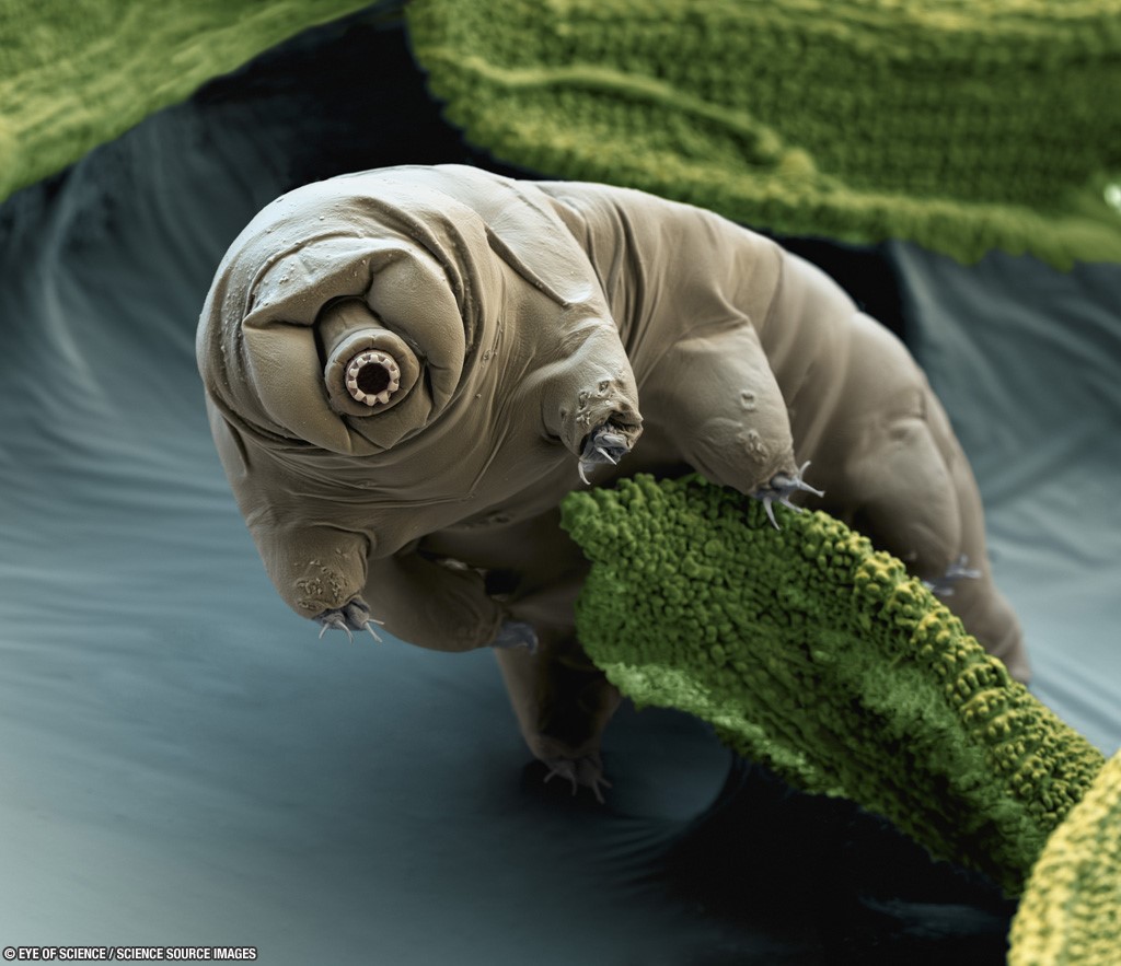
A breakthrough in studying tardigrades happened in 2016 when a genome of the most radiation-resistant species of tardigrades Ramazzottius varieornatus was sequenced by Japanese scientists. Comparing the genome of tardigrades to all known genomes of other organisms, a unique protein was found, named Damage suppressor, or Dsup for short.
Research on the impact of radiation, especially of cosmic radiation, on cognitive functions and morphological features of living organisms is being performed at the Joint Institute for Nuclear Research. The results are not encouraging, the impact is destructive, and it has already questioned the possibility of distant space flights. Geneticists hope that Dsup studies will provide them with a solution of how to reduce harmful consequences of radiation in the future. This protein may allow increasing the radio resistance level of an organism, or shielding tissues surrounding a tumour during radiotherapy, and serve as a DNA protector during long-lasting cryoconservation of samples of biological materials.
DLNP scientists are examining mechanisms of the Dsup protein to estimate the prospects for its application to increase radio resistance of complex multicellular organisms. A corresponding scientific project started at JINR last January. Earlier, studies of this kind were carried out in Japan, but only with human cultured cells HEK293 and X-rays. DLNP scientists opt for fruit flies Drosophila melanogaster and human cultured cells HEK293, a human embryonic kidney, as objects of research.
Thanks to the investigations in Japan, the gene sequence that codes the Dsup protein was already known, and the DLNP geneticists performed its chemical synthesis. After that, the gene was completely sequenced and optimised for using in research objects. “A microinjection of the solution containing the necessary DNA was made into Drosophila embryos,” says Elena Kravchenko, the Head of the DLNP Sector of Molecular Genetics of the Cell. “Adults developed from these embryos with the necessary genetic insertion segment to be now inherited by next generations.” After the procedure, specialists get a large fruit fly population with the inserted Dsup gene, which synthesises the Dsup protein in each cell of an organism.
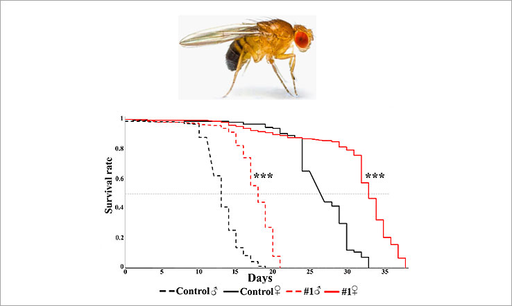 Figure 1: results of irradiation of strains of D. Melanogaster expressing Dsup and control groups. Curves display the survivability of individuals after irradiation. Black curves show control strains, red curves indicate the strains expressing Dsup
To find out how new Drosophila strains withstand radiation, samples were irradiated with gamma-quanta at an energy of 1―10 MeV with a dose of 500 Gy at the Microtron MT-25 accelerator in the Flerov Laboratory of Nuclear Reactions JINR. The results demonstrate an increase in radio resistance in the Drosophilae with the Dsup protein inserted into DNA (Figure 1).
HEK293 cells were irradiated with protons with a dose of 4 Gy at an energy of 150 MeV at the DLNP Phasotron. Figure 2 displays that the survivability of cells expressing Dsup is considerably higher than of those of the control strain.
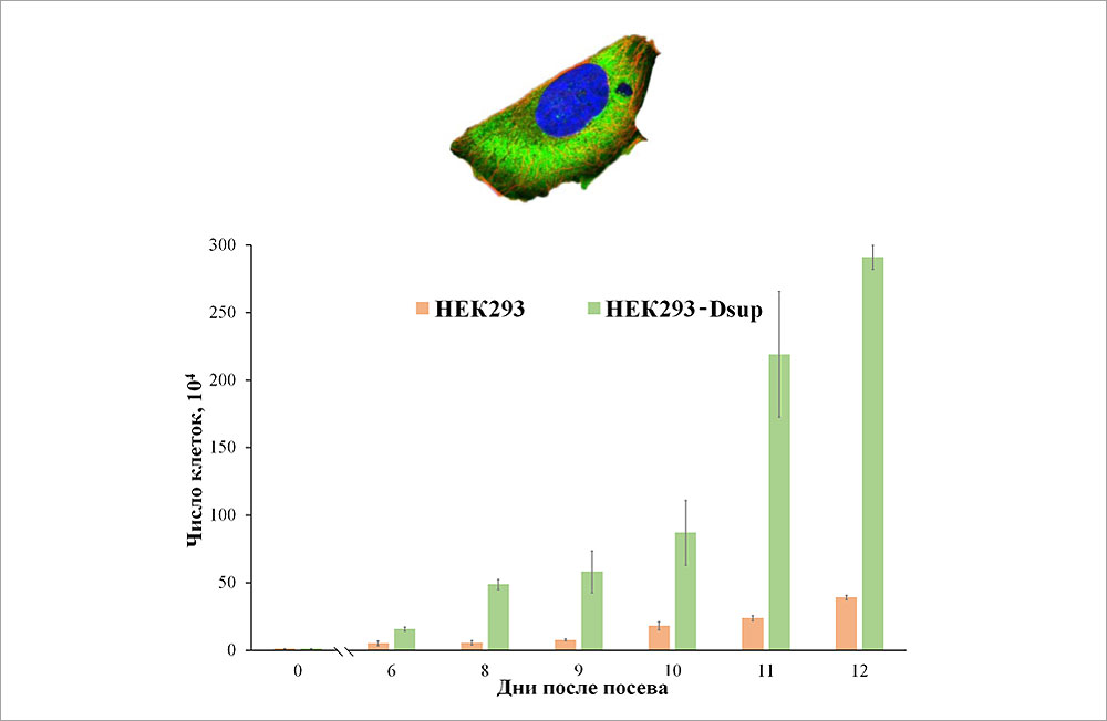 Figure 2: results of irradiation of HEK293 cells expressing Dsup and of control groups
Thus, JINR scientists have proved that the Dsup protein works in DNA of both fruit flies and human cultured cells HEK293, which means it is versatile. The study also demonstrated that radio resistance increases with exposure to different radiation types.
However, it is still obscure how this protein affects other systems of an organism. To reveal this, DLNP scientists performed a transcriptome analysis, in other words, tested activities of all genes involved in all biological processes of a fruit fly.
It turned out that more than 1,000 genes involved in crucial processes of an organism changed their activity. For example, activity of the genes involved in chromatin organization and transcription regulation, nervous system functioning, and others was reduced, i. e. the activity of processes related to the “DNA service” and information readout slowed down. It means that the way to avoid these negative consequences of Dsup use should be found. According to DLNP scientists, this gene constantly operates very actively during the whole life of a fruit fly. That is why a possible solution of the problem may become the turn-on and turn-off function of this gene. To do this, the Dsup gene activity should be adjusted so that as to turn it on and off by adding a particular substance, and also to operate only in appropriate time, e. g., during a space flight. One of the next objectives of this research team is to probe this hypothesis.
The research team is determining the Dsup protein structure to understand its operating mechanism. This objective required synthesising Dsup in large quantities, and it took several months until scientists from DLNP and FLNP fulfilled that task. As a result, they managed to get the insight into the Dsup secondary structure (three-dimensional form of protein local segments) using such methods as small-angle X-ray scattering (SAXS, research performed at FLNP JINR, MIPT, Moscow, and ESRF, Grenoble, France), small-angle neutron scattering (SANS), dynamic light scattering (DLS) (study conducted at FLNP). Thus, the protein size has been found (approximately 200 by 66 angstroms). Moreover, it has been proved that it is not globular (i. e. polypeptide chains are not folded into globules), and the existence of a secondary structure is established. To specify the Dsup secondary structure, scientists should conduct measurements using circular dichroism methods, DLS, and SAXS in native and denaturing conditions when structural failure is examined under the impact of different factors.
Experiments of this kind have been already scheduled for 2023 at the facilities of FLNP (XEUSS 3.0) and MIPT (J-1100 CD Spectrometer). One more objective will be to estimate the metabolic activity and induction of apoptosis (programmed cell death) in human cultured cells HEK293 expressing Dsup, as well as to determine the cellular oxygen level.