Transfer of JINR Expertise to Member States
S. E. Kichanov, E. V. Lukin, D. P. Kozlenko, V. N. Shvetsov, S. A. Kulikov, B. A. Abdurakhimov, M. Yu. Tashmetov, B. S. Yuldashev, N. B. Ismatov, A. R. Saidov, A. Normurodov
Development of neutron scattering instrumentation based on reconstructed or updated nuclear reactors is ongoing trend in several countries. The parameters of such nuclear research reactors are potentially sufficient for experimental studies of condensed matter by neutron scattering methods in the interdisciplinary fields, including condensed matter physics, materials science, non-destructive structural diagnostics, cultural and natural heritage, etc. [1, 2].
One of relatively simple and effective neutron scattering techniques is the neutron imaging (radiography and tomography). At the FLNP JINR, the proficient expertise in research using neutron imaging methods and development of relevant instrumentation have been gained during the recent years [3]. This expertise has become a subject of specific interest from the JINR Member states, having its own old reactors, recently renewed and required development of neutron scattering instrumentation. A good example is the neutron radiography and tomography facility, which was developed on the WWR-K research reactor in the Institute of Nuclear Physics (Almaty, Republic of Kazakhstan) jointly by the Frank Laboratory of Neutron Physics of JINR and INP staff [4].
Recently, the Directorate of the Institute of Nuclear Physics of the Academy of Sciences of the Republic of Uzbekistan (INP ASRUz) expressed interest in collaboration with FLNP JINR for development of the neutron imaging facility at the WWR-SM reactor. The WWR-SM research reactor, a representative of Water-cooled Water-moderated type Reactors, is located in the Ulugbek settlement of 30 kilometers from Tashkent. It resumed operation in 2017 and now this reactor is used in a wide range of scientific areas like nuclear physics, neutron capture therapy, neutron activation analysis and irradiation of minerals, as well as for the production of radioisotopes for nuclear medicine. Taking into account growing trend of requests from the scientific community in interdisciplinary applied studies [2, 3] in engineering and plant science, geophysics, astrophysics, archeology and paleontology, it was decided to develop a new neutron imaging experimental facility. In a relatively short time, the design of the new facility, ordering and construction of necessary equipment and the installation of main components were performed by joint JINR-INP ASRUz working team. Finally, the neutron imaging facility was put into operation in 2020.
 Fig. 1. The layout of the neutron imaging facility at the 5th beamline of the WWR-SM reactor [5]. The collimator system in the evacuated tube (1), the concrete biological shielding (2), detector system and goniometer position (3) are shown. The rotation goniometer (4), the scintillation screen position (5), and light-tight boron-contained polyethylene box with CCD-camera and optical lens are shown in photos of the detector system of the neutron imaging facility
The neutron beam is formed using a composite collimation system, which consists of several layers: the paraffin part with the length of 300 mm, boron-contained polyethylene of 100 mm, cadmium foil of 1 mm, and lead layer with the dimension of 100 mm. It provides protection against both gamma radiation and fast neutrons, which are present in the incident neutron spectrum. The characteristic parameter of the L/D ratio, characterizing neutron beam divergence of the facility, is 600. The detector system of the facility is shown in Figure 1. The scintillation screen 6LiF/ Zn(Cd)S:Ag manufactured by RC TRITEC Ltd (Switzerland) is used as a neutron converter. Thickness of the scintillator is 0.2 mm. The light is reflected out of the beam by two mirrors and focused on CCD chip of the camera ProLine PL-09000 manufactured by Finger Lakes Instrumentation (New York, USA). The spatial resolution of the neutron imaging facility is 280 μm.
As an example of the first neutron radiography experiments on the new facility, the neutron image of a metal casing padlock is shown in Figure 2. The inner lock gears and steel arc are visible and well distinguishable. There is a good neutron contrast between the different parts of the metal lock. Also, the neutron radiographic image of the complex system of the corn roots are shown in Figure 3. The neutrons easily penetrate through a plastic container and provide visual data about hidden organic matter.
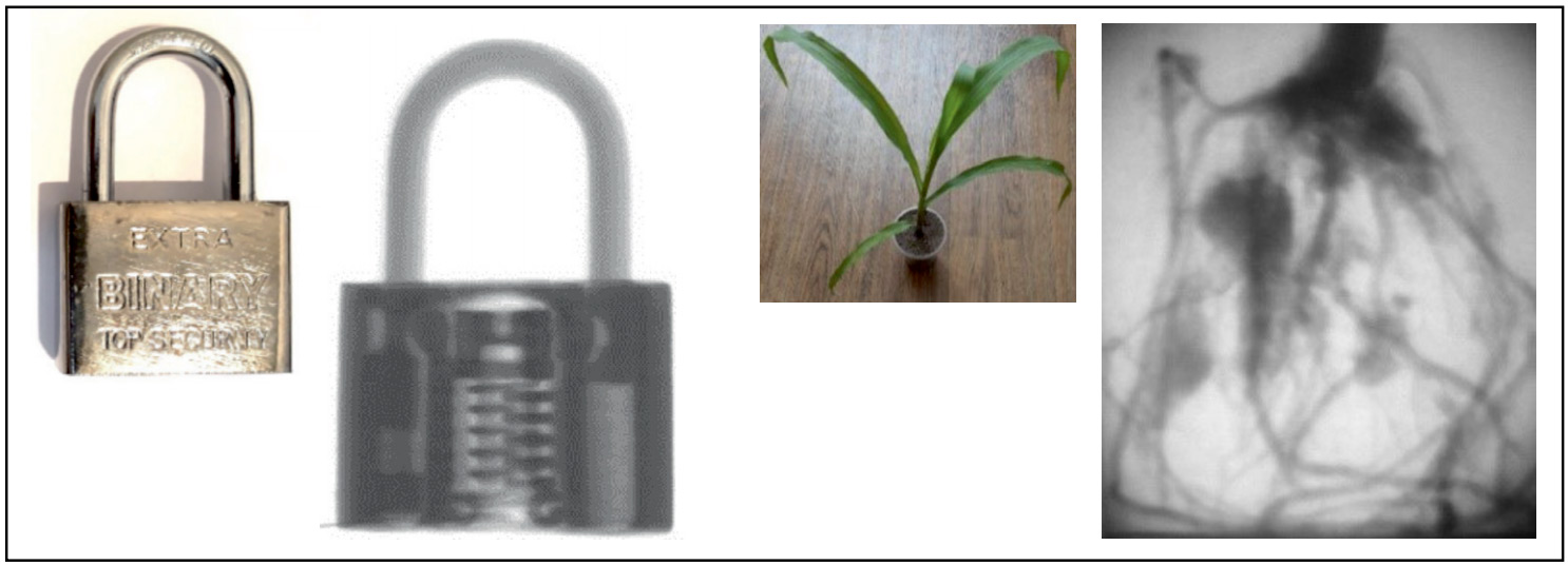 Fig. 2. The photo and neutron imaging of the metal padlock. The photo of the corn and the neutron radiographic image of the roots of the corn plant in a plastic container
The ancient Uzbekistan cultural place was an important trading center on the Silk Route [6], and the Uzbek nation is a rich mixture of the different cultures, as a result of the clash of civilizations. The non-destructive character of the neutron radiography and tomography method has prompted the rising interest in studying rare archaeological items. One of the important research directions that will be developed at the new neutron facility is the non-destructive neutron studies of the cultural heritage objects [2, 7]. The first studied cultural object was the bronze deer-shaped incense burner (Figure 3), dated to the III-IV centuries A.D. and found at an archeological site around the Dalvarzintepa settlement of the Surkhandarya region of the Uzbekistan. The inner arrangement of the empty space inside metal body of incense burner has been observed.
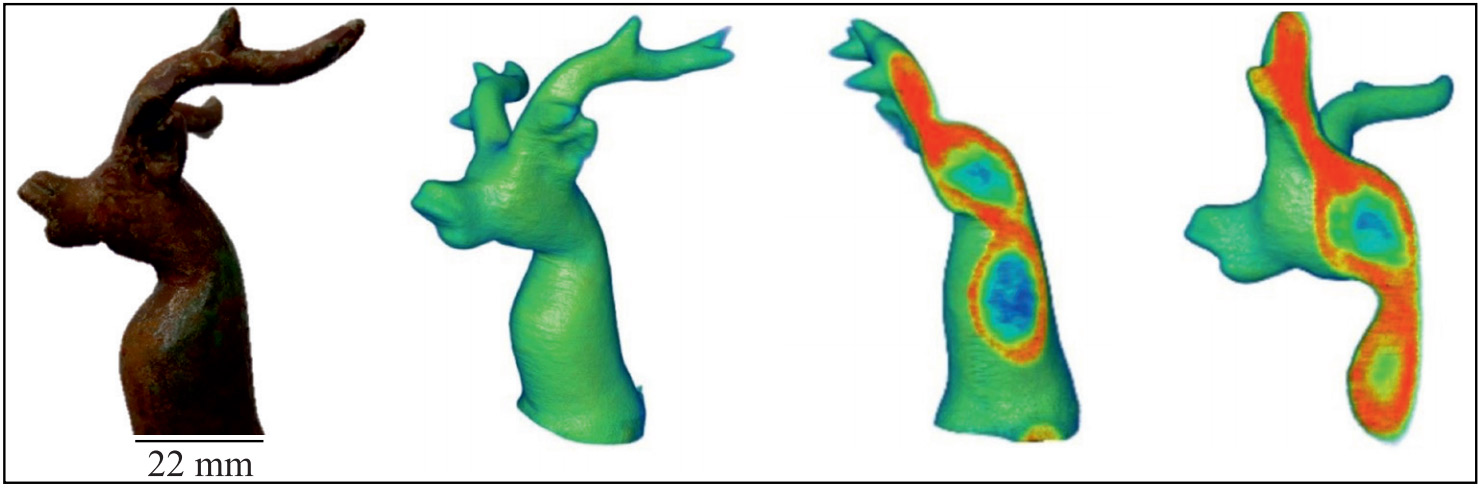 Fig. 3. Photo of bronze deer-shaped incense burner. The scale bars are shown. The 3D model after tomographic reconstruction procedure and different virtual slices of the obtained 3D data. The rainbow-like coloring shows the attenuation coefficients of the neutron beam from low (green) to high (red)
These first results demonstrate a significant potential for research in various multidisciplinary areas, including engineering and materials sciences, plant physiology, cultural and natural heritage sciences. The joint work provides an opportunity to bring cooperation between the institutions to a new level. Based on the experience of FLNP JINR, further activities will be focused on the improvement of the technical parameters and the realization of an extensive scientific program at the constructed neutron imaging facility.
References
- Lehmann E. H., Peetermans S., Betz B. Instrumentation in Neutron Imaging — A world-wide overview // Neutron News. 2015. V. 26. P. 6‒10.
- Podurets K. M., Kichanov S. E., Glazkov V. P., Kovalenko E. S., Murashev M. M., Kozlenko D. P., Lukin E. V., Yatsishina E. B. Modern Methods of Neutron Radiography and Tomography in Studies of the Internal Structure of Objects // Crystallography Reports. 2021. V. 66, No. 2. P. 254‒266.
- Kozlenko D. P., Kichanov S. E., Lukin E. V., Rutkauskas A. V., Belushkin A. V., Bokuchava G. D., Savenko B. N. Neutron Radiography and Tomography Facility at IBR-2 Reactor // Phys. Part. Nuclei Lett. 2016. V. 13. P. 346.
- Nazarov K. M., Muhametuly B., Kenzhin E. A., Kichanov S. E., Kozlenko D. P., Lukin E. V., Shaimerdenov A. A. New Neutron Radiography and Tomography Facility TITAN at the WWR-K Reactor // Nuc. Inst. and Meth. in Phy. Res. Sec. A. 2020. P. 164572.
- Abdurakhimov B. A., Tashmetov M. Yu., Yuldashev B. S., Kichanov S. E., Lukin E.V., Kozlenko D. P., Kulikov S. A., Shvetsov V. N., Ismatov N. B., Saidov A. R., Normurodov A. B., A. V. Rutkauskas. New Neutron Imaging Facility at the WWR-SM Reactor: Design and First Results // Nuc. Inst. and Meth. in Phy. Res. Sec. A. 2020. P. 164959.
- Kilic-Schubel N. Timurid Empire // The Encyclopedia of Empire. 2016. P. 1‒11.
- Kichanov S. E., Saprykina I. A., Kozlenko D. P., Nazarov K., Lukin E. V., Rutkauskas A.V., Savenko B. N. Studies of Ancient Russian Cultural Objects Using the Neutron Tomography Method // J. Imaging. 2018. V. 4, No. 2. P. 25.