Over the past few years, Raman spectroscopy has become a powerful diagnostic tool in the life sciences. Studies have been performed on the application of Raman spectroscopy for the distinction of neutrophils transformed during NETosis. Neutrophils are the most common human blood leukocytes, which are the most important part of the innate immunity and carry out a fast response to microbial and viral invasion.
The research was carried out by a group of scientists of the Sector of Raman Spectroscopy of the Frank Laboratory of Neutron Physics of JINR headed by Grigory Arzumanyan in the collaboration with scientists from Vladimirsky Moscow Regional Clinical and Research Institute, R&D Department of the Medtechnopark Ltd., Prokhorov General Physics Institute, Russian Academy of Sciences and Moscow Chemical Lyceum. The article was published in the 3rd issue of the bulletin “JINR News” 2020.
NETosis is a process of the programmed neutrophil cell death involved in the development of many diseases, including those associated with high mortality. During the NETosis process, neutrophils undergo cardinal transformation resulting in the destruction of the cell with the release of its internal contents. As a result of NETosis, the socalled Neutrophil Extracellular Traps (NETs) arise. NETs are composite DNA complexes with neutrophil proteins transformed in the NETosis process. Our goal was to search for possible spectral markers in neutrophil Raman spectra, caused precisely by NETotic transformation of neutrophils. From the point of view of spectroscopy, we were aimed at finding significant differences in the Raman spectra caused by the transformation of neutrophils.
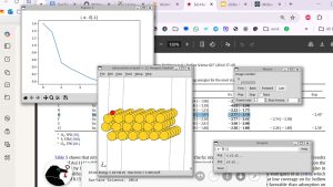
A confocal microspectroscopy setup with high spatial resolution was used for Raman measurements. It comprises a Confotec CARS scanning laser spectrometer (SOL Instruments Ltd., Belarus) coupled to the NIKON TE2000-E inverted microscope. At the initial stage of NETs formation under the action of special enzymes, the antimicrobial elements are citrullinated in the granules of the neutrophil cell, indicating the start of the NETosis activation process.
Highly sensitive Raman spectroscopy revealed in the low-frequency range the evolution (growth) of the citrulline peak within 30–40 min after the beginning of the inflammatory process, which can be classified as an early diagnosis of NETosis.
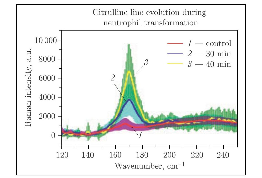 Low-frequency range of Raman spectra of neutrophils: citrulline line evolution (growth) indicating the pre-activation of NETosis
Arzumanyan G., Mamatkulov K., Volkov A., Vereschagin K. et al. // J. Raman Spectroscopy. 2020. V. 1. P.10; https://doi.org/10.1002/jrs.5844.