Scientists of the Frank Laboratory of Neutron Physics JINR within the joint research team are employing neutron scattering techniques to study the structure of wound dressings based on bacterial cellulose and gain an insight into the structural changes that occur during cellulose degradation by enzymes. Their findings can help to develop biodegradable dressings with the desired properties for the treatment of burns and other skin lesions.
Kombucha and coconut jelly, textiles, paper and sensitive acoustic membranes, biocompatible implants and versatile wound dressings – all these are about bacterial cellulose (BC), an amazing substance discovered in 1886, fifty years after the discovery of the well-known plant cellulose. Although mankind has been familiar with bacterial cellulose for a long time (Philippine desserts nata de coco and nata de piña), its properties began to be investigated only in the last century.
Cellulose produced by bacteria (for example, acetic acid bacteria Gluconoacetobacter xylinum) in the form of a gelled film with a fine-fiber net structure, has a smaller fiber diameter and an increased surface area compared to those in plant cellulose, and exhibits exceptional properties, including high mechanical strength and elasticity, water and vapor permeability, high water absorption capacity, non-toxicity and good biocompatibility. What is more, bacterial cellulose can promote wound healing. These unique physicochemical and biological properties have found application in medicine, and for over ten years they have been used to develop hemostatic agents, implants and artificial blood vessels, artificial skin tissue and skin wound dressings.
Wound dressings based on bacterial cellulose are becoming a promising alternative to traditional bandaging materials, but the requirements for wound dressings are quite extensive. They should be easy to apply and painlessly removed, be biocompatible, not cause allergic reactions, protect against bacterial infections and provide a moist environment and effective oxygen circulation. An additional important requirement is the ability to change shape at a rate matching the formation of new tissue. That is, BC-based wound dressings should have a controlled biodegradation rate, but there are no enzymes in the human body that are capable of catalyzing cellulose degradation.
To obtain a controlled technology based on enzymatic hydrolysis of cellulose, it is necessary not only to understand the relationships between the structure and the physical and chemical properties of biomaterials, but also to possess tools that can affect their changes.
As a variant of such a tool for improving the properties of bacterial cellulose for biomedical applications, scientists from PNPI, in a joint study with specialists from a number of research centres, used the fungal enzyme cellobiohydrolase isolated from yeast-like fungus Scytalidium candidum 3C, and for the first time studied the process of BC degradation in detail.
But what do neutrons have to do with it?
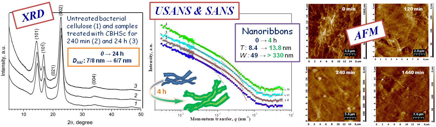
“In our study, we used a wide range of physical and microscopic techniques including small-angle neutron scattering, X-ray diffraction and atomic force microscopy (Dubna, FLNP JINR), ultra-small-angle neutron scattering (Garching, Germany, Heinz Maier-Leibnitz Zentrum), as well as scanning electron microscopy (Moscow, Center for Collective Use of Kurnakov Institute of General and Inorganic Chemistry of RAS). This allowed us to obtain comprehensive data on changes in the crystalline and supramolecular structure of cellulose nanogel films at a level from several nanometers to several micrometres at different stages of their hydrolysis during the enzymatic treatment,” explained Yulia Gorshkova, a researcher from the Frank Laboratory of Neutron Physics, JINR. “One of the reasons why bacterial cellulose is actively used in the engineering of materials for skin and bone regeneration is its morphological similarity to collagen, which is the main structural protein with a fibrillar structure found in the body’s various connective tissues. Fibrils of native collagen are larger than fibrils of native bacterial cellulose and have a cylindrical shape, while fibrils of native BC have a lamellar shape. Under the action of the enzyme, as shown by neutron scattering methods, the size and shape of the components of the BC polymer matrix begin to approach those in natural collagen.”
The possibility of safe application of enzyme-treated bacterial cellulose on biological systems was proved in experiments in vivo and in vitro. The experiments revealed no cytotoxic effects associated with the enzyme addition to BC dressings and showed a generally positive influence on the treatment of extensive III-degree burns, significantly accelerating wound healing.
The obtained results will contribute to the creation of biotechnologies for the development of wound dressings with desired properties for the treatment of various skin lesions.
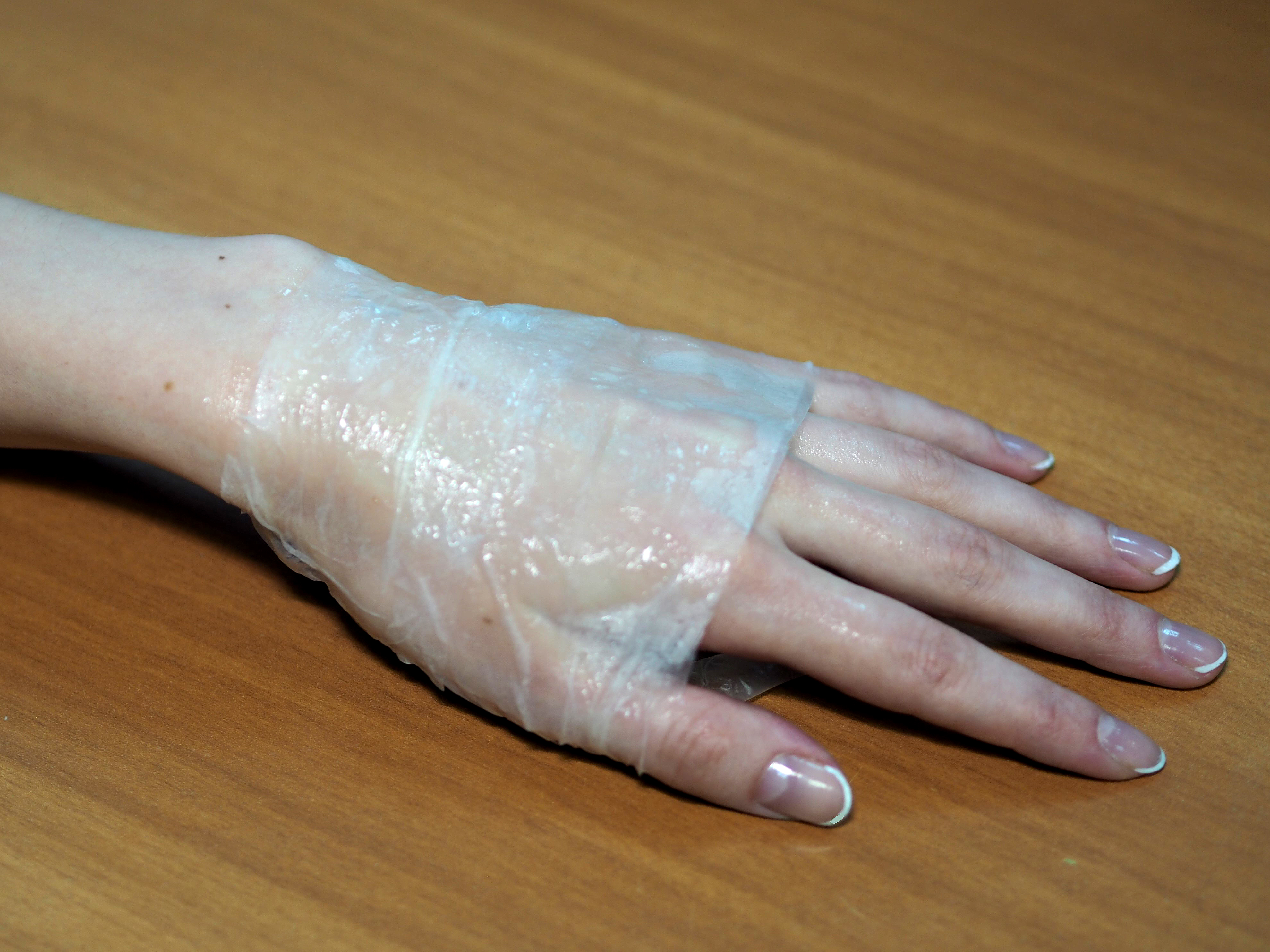
 Structural changes in BC as a result of its treatment with enzyme
Published in Materials:
L. Ivanova etc. Crystal and Supramolecular Structure of Bacterial Cellulose Hydrolyzed by Cellobiohydrolase from Scytalidium Candidum 3C: A Basis for Development of Biodegradable Wound Dressings. Materials 2020, 13, 2087; doi:10.3390/ma13092087
This article is an open access article distributed under the terms and conditions of the Creative Commons Attribution (CC BY) license (http://creativecommons.org/licenses/by/4.0/).
Olga Baklitskaya-Kameneva