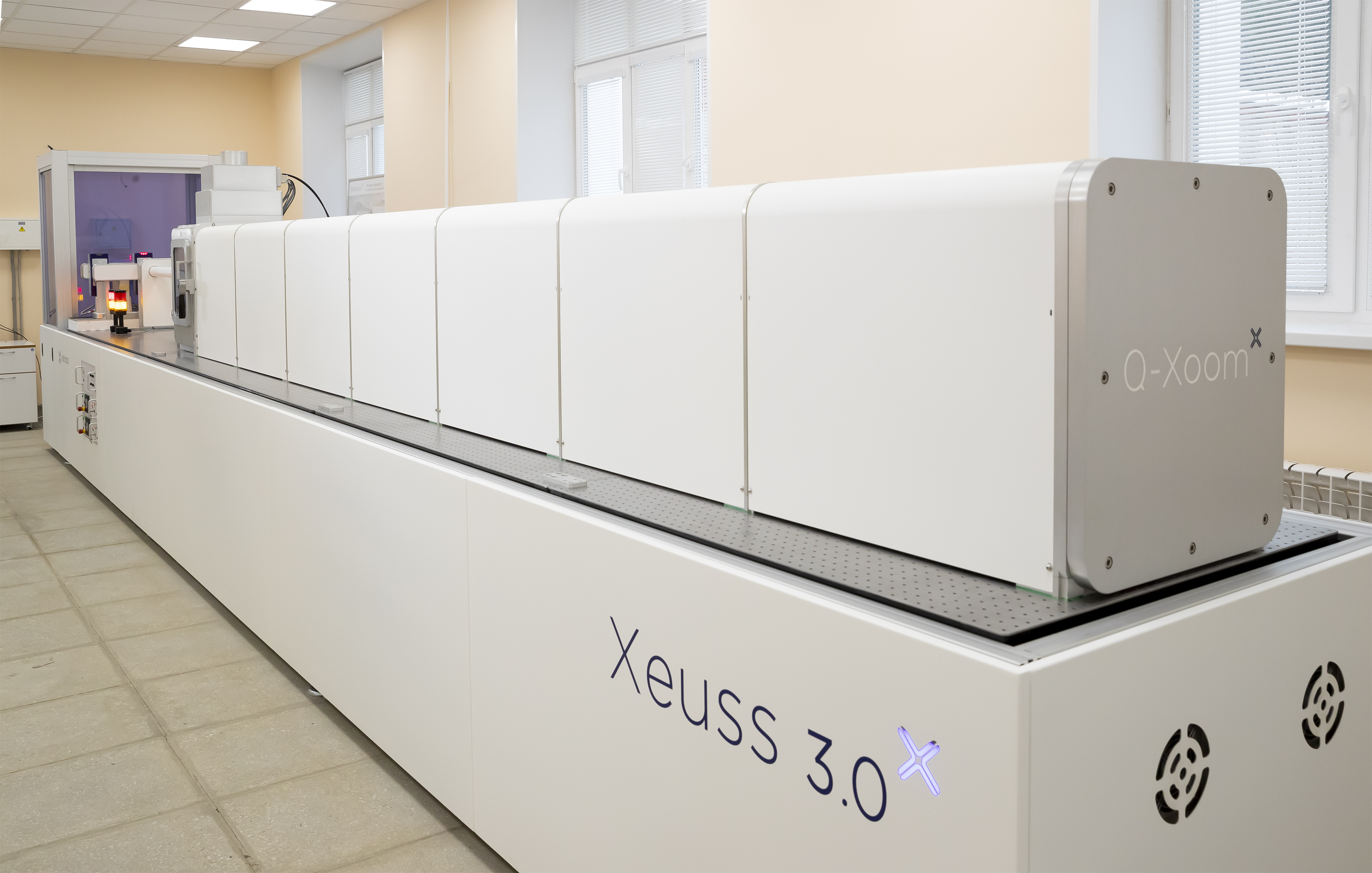
Modern research is hard to imagine without life science investigations. Scientists strive not only to learn the fundamental laws of the Universe, but also to improve the quality of human life, solve acute problems of healthcare, agriculture, ecology and find new sources of energy. Life science investigations have long and firmly taken their place among research topics of the Joint Institute for Nuclear Research. JINR has launched the creation of the MSC-230 medical cyclotron; research on the effect of cosmic radiation on living organisms, as well as genetic studies of longevity are underway; a vast amount of experience has been gained in the field of proton therapy; the ARIADNA complex for applied research, including medical research, is being actively developed at the future NICA collider. And this list is far from complete. For these purposes, the suite of instruments of the Institute is constantly being expanded. The new Xeuss 3.0 X-ray scattering station (FLNP JINR) is also used, among other things, for research in this field of knowledge.
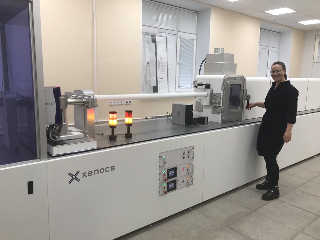
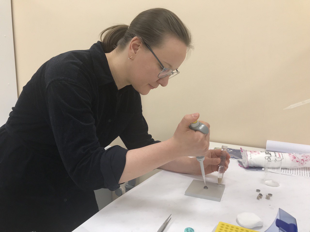
A rich variety of sample holders allows specialists to study biological objects in solution. Such investigations were recently carried out by Ekaterina Iashina, a researcher from the B.P. Konstantinov St. Petersburg Nuclear Physics Institute (PNPI), PhD in Physics and Mathematics, who defended her dissertation at JINR in 2020.
“The main object of our research is chromatin, the contents of the nucleus of a biological cell. The nucleus stores DNA that carries genetic information for the development and functioning of an organism. It is a very large molecule: if you expand the human DNA, its length will be about two meters,” Ekaterina said. This long polymer molecule is ‘packed’ in the nucleus with the help of special architectural proteins, i.e., it is folded in a certain way in the nucleus. An interesting fact is that the volume of the nucleus is relatively small, but at the same time, such a structure still allows performing all biologically inherent functions. “The molecule cannot be arranged chaotically, like a thread in a ball,” the researcher emphasized. — There is a special multilevel organization of chromatin. This is what we are studying with the help of small-angle scattering at the Xeuss 3.0 station in FLNP.”
Most of the biological discoveries in the field of DNA research have been made using the microscopy method. However, it has a number of limitations. For example, it allows one to see only the surface of the sample under study, and in addition, it is not always possible to study the object in native conditions, as well as there are restrictions on the sample size. Therefore, scattering methods are widely used to study the internal structure of biological objects.
The main method for studying chromatin in the nucleus of a biological cell, which is employed by the scientific group from PNPI under the direction of Sergei Grigoriev, in which Ekaterina works, is small-angle and ultra-small-angle neutron scattering. During the five years of their work, the nuclei of chicken erythrocytes, HeLa cells and rat lymphocytes were studied using the KWS-2 and KWS-3 diffractometers of the International Heinz Maier-Leibnitz Zentrum, Germany, Garching. Due to technical problems, the work of the Center has been suspended for the past two years, which prompted the scientists to master new methods and new experimental stations.
“This is my first experience with X-rays. Since there is a shortage of neutrons now, we are exploring new horizons, new methods,” Ekaterina emphasized. She noted a number of advantages of the X-ray instrument for the study of biological objects. Since the intensity of X-ray and synchrotron sources is significantly higher than that of neutron sources, measurements are performed with a higher resolution, due to which structural features of the object under study are reflected better in the scattering curve, measurements are conducted faster, and a much smaller sample volume is required. For experiments at Xeuss 3.0, the required sample size is several times smaller than for experiments on the best small-angle neutron station, and the data acquisition rate is approximately twice as high. However, the contrast variation technique, which is widely used in biological research and has been well tested in neutron scattering, is still difficult to employ in X-ray scattering experiments, although it is possible.
According to Ekaterina Iashina, the DNA molecule is packed in a fractal way. “Prior to our studies on Xeuss 3.0, in small-angle neutron scattering experiments in Germany, we found that chromatin has a bi-fractal structure,” Ekaterina said. “On a large scale, the structure of chromatin is described by a logarithmic fractal model, and on a smaller scale, by a mass fractal model.”
The term ‘fractal’ was first introduced by Benoît Mandelbrot in the late 1960s of the last century. It is used to describe complex objects. In fact, this is a kind of self-similarity principle, in which one structural element of an object is repeated in the next. The most striking examples of fractals are the Sierpiński triangle, the Koch snowflake, or any coastline. All of the above examples correspond to regular fractals, which, within the framework of the concept of small-angle scattering by fractals, correspond to mass fractals, which are characterized by corresponding fractal dimensions. The term ‘logarithmic fractal’ was introduced relatively recently, and one of the many examples of a logarithmic fractal is a fractal describing the area-preserving rule for botanical trees by Leonardo da Vinci [1].
So far, the group of scientists from PNPI has succeeded in studying several samples. To study the structure of chromatin using neutrons, Ekaterina Iashina and her colleagues used the nuclei of chicken erythrocytes [2], the cell line of HeLa nuclei (cancer cells) [3], and the nuclei of rat lymphocytes (immune cells) [4]. All three samples showed a bi-fractal organization of chromatin with mass and logarithmic fractals. The scientists believe that such a bi-fractal organization of chromatin in the nuclei of biological cells is universal and necessary for the cell to perform its biological functions. However, this hypothesis needs to be tested by numerous experiments and simulations. It is also important to understand what can affect chromatin, and how we can change its structure. For this purpose, the researchers conducted several experiments to understand how the structure of chromatin is related to the biological functions of DNA, such as transcription, that is, reading information about the structure of proteins that the cell should synthesize, or DNA repair, that is, correcting damage in DNA molecules. Using the Xeuss 3.0 station, the researchers from PNPI study the nuclei of immortalized cell lines treated with special substances, and explore how enhanced repair or transcription suppression changes the structure of chromatin.
- S. V. Grigoriev, O. D. Shnyrkov, P. M. Pustovoit, E. G. Iashina, and K. A. Pshenichnyi, Experimental evidence for logarithmic fractal structure of botanical trees, Phys. Rev. E 105, 044412 (2022)
- S. V. Grigoriev, E. G. Iashina, V. Yu. Bairamukov, V. Pipich, A. Radulescu, M. V. Filatov, R. A. Pantina, and E. Yu. Varfolomeeva, Switch of fractal properties of DNA in chicken erythrocytes nuclei by mechanical stress, Phys. Rev. E 102, 032415 (2020)
- S. V. Grigoriev, E. G. Iashina, B. Wu, V. Pipich, Ch. Lang, A. Radulescu, V. Yu. Bairamukov, M. V. Filatov, R. A. Pantina, and E. Yu. Varfolomeeva, Observation of nucleic acid and protein correlation in chromatin of HeLa nuclei using small-angle neutron scattering with D2O-H2O contrast variation, Phys. Rev. E 104, 044404 (2021)
- E. G. Iashina, E. Yu. Varfolomeeva, R. A. Pantina, V. Yu. Bairamukov, R. A. Kovalev, N. D. Fedorova, V. Pipich, A. Radulescu, and S. V. Grigoriev, Bifractal structure of chromatin in rat lymphocyte nuclei, Phys. Rev. E 104, 064409 (2021)