The second JINR Prize for 2019 in the section “Applied research and technology papers” was awarded to the team of authors comprising of D. P. Kozlenko, S. E. Kichanov, A. V. Belushkin, E. V. Lukin, K. Nazarov, A. V. Rutkauskas, G. D. Bokuchava, B. N. Savenko, I. A. Saprykina was awarded for the scientific paper “Neutron radiography and tomography at the pulsed high-flux IBR-2 reactor: development of the experimental facility and results of the interdisciplinary applied research”.
The neutron radiography and tomography methods, providing possibilities to obtain the images and three-dimensional reconstructed models of the internal structure of studied objects with a spatial resolution of about 100 µm, are now extensively developed at the modern neutron sources. The high penetration depth of neutrons gives certain advantages to these methods in comparison with relevant X-ray ones and such methods can be effectively used for interdisciplinary research in the wide range of topics: from the studies of the structural features of various functional materials, the microstructure of building and construction materials and their changes during ongoing processes, to investigations of the internal organization of unique cultural and natural heritage objects and the extraterrestrial objects.
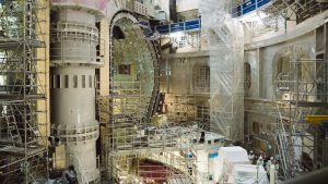
Not so long ago, the neutron radiography and tomography facility have been developed at the Frank Laboratory of Neutron Physics JINR on the 14th beamline of the IBR-2 high-flux pulsed reactor, and activities were made to improve its technical parameters. The development of this facility (fig. 1) has stimulated the evolution of a new applied research direction, related to non–destructive analysis of the internal organization of a wide range of objects and materials, including paleontological representatives of natural heritage [1], engineering products [1, 2, 5], the objects of extraterrestrial origin – meteorites [1, 6], archaeological objects of cultural heritage [7-11].
 Fig. 1. Top: the layout of the neutron radiography and tomography facility on the 14th beamline of the IBR-2 high-flux pulsed reactor. The length of the collimation system (1) is 11 m, and the linear dimensions of the field of view at the sample object position are 20×20 cm. The goniometer system (2) is used for the neutron tomography experiments. A specially constructed detector system (3) based on a high-resolution CCD camera is used for recording of neutron images. Bottom: a principal scheme of the neutron radiography experiment: a neutron image of the object is formed using the scintillator screen. The neutron radiography image is recorded by means of the CCD video camera.
The performed experimental studies enabled to reconstruct the internal structure of the fossil cone Protosequoia sp. (fig. 2), dated by the late Cretaceous period (around 100 million years ago). The reconstructed three-dimensional model of the cone stem and petals was obtained [1]. Also, the volume distribution of a Cyanobacteria colony inside a fossil stromatolite was studied [1]. Colonies of these ancient bacteria provide information about the evolution of living organisms on the Earth more than 3.5 billion years ago. The obtained neutron experimental data is important for the development of models of the evolution of these organisms.
 Fig. 2. Photo (left) and the reconstructed from the neutron tomography data three-dimensional model of the internal structure of a fossilized cone Protosequoia sp. The remains of the damaged stem of the cone and the arrangement of cone petals are clearly observed on the three-dimensional model of the studied object.
Three-dimensional models of the internal structure of the meteorites Seymchan (fig. 3) [6] and Marjalahti [1] have been obtained using the neutron tomography method. The peculiar features of the neutron interactions with matter allowed us to detect and separate the internal mineral and metallic components of these meteorites, to obtain a three-dimensional spatial distribution of nickel in the metallic components of these meteorites-pallasites [1, 6], and to determine distributions of the volumes and average sizes of internal composite minerals, as well as their morphological features [6].
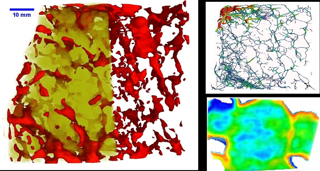 Fig. 3. The three-dimensional model of the Seymchan meteorite, which is reconstructed from neutron tomography data. The red areas correspond to a metallic iron-nickel alloy component, and the yellow one to an olivine mineral component. To obtain quantitative characteristics of the metal component, a skeletal dendritic structure was used in the analysis of three-dimensional data (top right). This skeletal structure reflects the distribution of the metal alloy in the meteorite volume and allows to build distributions both along the length and thickness of the metal component in a particular point of the meteorite. In the lower right part of the figure, a slice of a three-dimensional model of a single grain of olivine in the Seymchan meteorite is presented. Features of neutron radiographic contrast formation allow us to observe the distribution of iron-containing minerals within the volume of each olivine grain.
An example of research in the field of engineering sciences is the study of the kinetics of melting of the mixture of ice and granulated quartz [5], which allowed us to study the dependence of the melting temperature of the mixture on the size of quartz granules. The obtained experimental information is of great interest in the improvement of the technology of cleaning of snow cover in the cold regions of the Earth.
The significant outcome of neutron radiography and tomography application for non-destructive investigations of cultural heritage objects should be particularly noted [7-11]. This includes unique studies of the internal organization and phase composition of the coins of Ancient Greece [7] and Ancient Bulgaria (fig. 4) [8], the items of Ancient Russian cultural heritage [9, 10], included ones from the “Tver Treasure” (figs. 5, 6) found in 2014 and dated to the XIII century of the period of the invasion of Batu Khan [11]. High neutron radiographic contrast between silver and copper provides a possibility for the successful investigations of the spatial distribution of content elements and evaluation of the chemical composition of ancient coins: from Bosporus stators [7] to ancient Bulgar pools [8].
 Fig. 4. Photo of the Samanid multidirham as an example of the Ancient Volga Bulgaria coins, dated to the time of Khan al-Amir as-said Abu Salih Mansur I bin Nuh, the first half of the X century (top left). A three-dimensional model of the studied multidirham reconstructed from neutron tomography data is presented. The highlighted areas in the coin volume are regions with enhanced silver content. Several transverse virtual slices of the reconstructed three-dimensional model of the multidirham are shown. The red areas correspond to regions of the coin with high silver content.
 Fig. 5. Photo (left) and the reconstructed from neutron tomography data three – dimensional model of the ancient Russian jewelry (ray colt) from the Tver treasure. Both the surface decoration elements and the internal components of the fastener were observed from the neutron tomography data.
 Fig. 6. Photo (left) and the reconstructed three-dimensional model of the fragment of the wide double-leaf bracelet from the Tver treasure. The interior decoration elements of the cultural heritage object were reconstructed using the neutron tomography method.
The high penetration ability of neutrons allows us to reconstruct the 3D internal structure of the ancient Russian [9, 11] and medieval European jewellery [10], to observe their hidden decorative elements, and to clarify the historical and cultural origin of these archaeological finds. The obtained experimental information is important for the restoration of these cultural heritage items, as well as for the elucidation of the ancient technologies for coinage, jewellery, and household items production.
The experimental capabilities of new neutron radiography and tomography facility, as well as the recently obtained results have already provoked great interest among the research community of JINR Member States. As a result, the neutron radiography and tomography facility has been developed at the WWR-K reactor (INP, Almaty) in the Republic of Kazakhstan with the participation of the FLNP JINR specialists. The first experimental results were obtained recently.
References
[1]. D. P. Kozlenko, S. E. Kichanov, E. V. Lukin, A. V. Rutkauskas, G. D. Bokuchava, B. N. Savenko, A. V. Pakhnevich, A.Yu. Rozanov “Neutron Radiography Facility at IBR-2 High Flux Pulsed Reactor: First Results” Physics Procedia, 69, 87-91 (2015).
[2]. D. P. Kozlenko, S. E. Kichanov, E. V. Lukin, A. V. Rutkauskas, A. V. Belushkin, G. D. Bokuchava, B. N. Savenko “Neutron radiography and tomography facility at IBR-2 reactor”, Physics of Particles and Nuclei Letters, 13, 3, 346-351 (2016)
[3]. A. V. Rutkauskas, D. P. Kozlenko, S. E. Kichanov, G. D. Bokuchava, E. V. Lukin, B. N. Savenko “Investigation of the Neutron Transmission Spectra of Materials Promising for the Manufacturing of Crystalline and Polycrystalline Filters” Journal of Surface Investigation X-ray Synchrotron and Neutron Techniques, 9(2): 317-319 (2015)
[4]. E. V. Lukin, D. P. Kozlenko, S. E. Kichanov, A. V. Rutkauskas, G. D. Bokuchava, B.N. Savenko “First attempts on energy-selective neutron imaging at IBR-2”, Physics Procedia, 69, 271-274 (2015)
[5]. L. Kalvoda, S. E.Kichanov, M. Kučeráková, E. V. Lukin, S. Vratislav “Ice Melting Kinetics in Sand–Water Mixtures Investigated by Neutron Radiography and Diffraction”, Journal of Cold Regions Engineering, 33, 3, 04019003 (2019)
[6]. S. E. Kichanov, D. P. Kozlenko, E. V. Lukin, A. V. Rutkauskas, E. A. Krasavin, A. Y. Rozanov, B. N. Savenko “A neutron tomography study of the Seymchan pallasite”, Meteoritics & Planetary Science, 53, 10, 2155-2164 (2018)
[7]. M. G. Abramson, I. A. Saprykina, S. E. Kichanov, D. P. Kozlenko, K. M. Nazarov “A Study of the Chemical Composition of the 3rd Century AD Bosporan Billon Staters by XRF-Analysis, Neutron Tomography and Diffraction”, Journal of Surface Investigation: X-ray, Synchrotron and Neutron Techniques, 12, 1, 114-117 (2018)
[8]. S. E. Kichanov, K. M. Nazarov, D. P. Kozlenko, I. A. Saprykina, E. V. Lukin, B. N. Savenko, “Analysis of the internal structure of ancient copper coins by neutron tomography”, Journal of Surface Investigation: X-ray, Synchrotron and Neutron Techniques, 11, 3, 585-589 (2017)
[9]. [9]. I. A. Saprykina, S. E. Kichanov, D. P. Kozlenko «Possibilities, Limitations, and Prospects of Using Neutron Tomography and Radiography for Preservation of Archaeological Heritage Objects», Crystallography Reports, 64, 1, 152-155 (2019)
[10]. S. E. Kichanov, I. A. Saprykina, D. P. Kozlenko, K. Nazarov, E. V. Lukin, A. V. Rutkauskas, B. N. Savenko, Studies of Ancient Russian Cultural Objects Using the Neutron Tomography Method, J. Imaging, 4(2), 25 (2018)
[11]. [11]. I. A. Saprykina, S. E. Kichanov, D. P. Kozlenko, E. V. Lukin “ The capabilities of neutron tomography in archaeology on an example of the study of Old Russian jewelry from the Tver hoard of 2014”, Russian Archeology, №3 (2018) 36-42